Mouse Secondary Antibodies
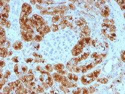
Biotium GP2 (Glycoprotein 2) / ZAP75 (GP2/1805), CF740 conjugate, 0.1mg/mL
GP2 (glycoprotein 2), also known as ZAP75, is a 537 amino acid secreted protein. It is an integral membrane protein that is secreted from intracellular zymogen granules and associates with the plasma membrane via glycosylphosphatidylinositol (GPI) linkage. GP2 is cleaved and then released into the pancreatic duct along with exocrine secretions. GP2 binds pathogens such as enterobacteria, thereby playing an important role in the innate immune response. The C-terminus of this protein is related to the C-terminus of the protein encoded by the neighboring gene, uromodulin (UMOD). GP2 is also expressed on the apical plasma membrane of specialized microfold (M) cells among enterocytes and serves as a transcytotic receptor for mucosal antigens. M cells are considered a promising target for oral vaccination against various infectious diseases.Primary antibodies are available purified, or with a selection of fluorescent CF Dyes and other labels. CF Dyes offer exceptional brightness


Biotium Desmin (Muscle Cell Marker)(D33), CF740 conjugate, 0.1mg/mL
Cytoskeletal intermediate filaments (IFs) constitute a diverse group of proteins that are expressed in a highly tissue-specific manner. IFs are constructed from two-chain -helical coiled-coil molecules arranged on an imperfect helical lattice, and have been widely used as markers for distinguishing individual cell types within a tissue and identifying the origins of metastatic tumors. Vimentin is an IF general marker of cells originating in the mesenchyme. Vimentin and Desmin, a related class III IF, are both expressed during skeletal muscle development. Desmin, a 469 amino acid protein found near the Z line in sarcomeres, is expressed more frequently in adult differentiated state tissues. Anti-desmin detects cells of normal smooth, skeletal, and cardiac muscles. Antibody reacts with leiomyomas, leiomyosarcoma, rhabdomyomas, rhabdomyosarcoma, and perivascular cells of glomus tumors of the skin. Primary antibodies are available purified, or with a selection of fluorescent CF
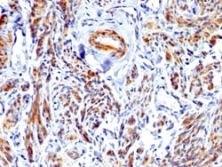
Biotium Caldesmon, HMW (h-Caldesmon) (Smooth Muscle Marker), CF568 conjugate, 0.1mg/mL
Caldesmon HMW is the high molecular weight variant of Caldesmon. Two closely related variants of human caldesmon have been identified which are different in their electrophoretic mobility and cellular distribution. The h-caldesmon variant (120'150 kDa) is predominantly expressed in smooth muscle whereas l-caldesmon (70'80 kDa) is found in non- muscle tissue and cells. Neither of the two variants has been detected in skeletal muscle. This antibody recognizes only the 150 kDa variant (h-caldesmon) in Western blots of human aortic media extracts and is unreactive with fibroblast extracts from cultivated human foreskin. Caldesmon is a developmentally regulated protein involved in smooth muscle and non-muscle contraction. Primary antibodies are available purified, or with a selection of fluorescent CF Dyes and other labels. CF Dyes offer exceptional brightness and photostability. Note: Conjugates of blue fluorescent dyes like CF405S and CF405M are not recommended for detectin
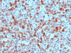
Biotium CD10 (Membrane Metalloendopeptidase) (MME/1893), CF568 conjugate, 0.1mg/mL
CD10 (also known as Common Acute Lymphocytic Leukemia Antigen (CALLA)) is a type II transmembrane protein. It is a cell surface enzyme with neutral metalloendopeptidase activity, which inactivates a variety of biologically active peptides. CD10 is expressed on the cells of lymphoblastic, Burkitt's, and follicular germinal center lymphomas, and on cells from patients with chronic myelocytic leukemia (CML). It is also expressed on the surface of normal early lymphoid progenitor cells, immature B cells within adult bone marrow and germinal center B cells within lymphoid tissue.Primary antibodies are available purified, or with a selection of fluorescent CF Dyes and other labels. CF Dyes offer exceptional brightness and photostability. Note: Conjugates of blue fluorescent dyes like CF405S and CF405M are not recommended for detecting low abundance targets, because blue dyes have lower fluorescence and can give higher non-specific background than other dye colors.
Biotium MUC1 / CA15-3 / EMA / CD227 (Epithelial Marker) (MUC1/1887R), CF740 conjugate, 0.1mg/mL
This MAb reacts with MUC1, a large transmembrane glycoprotein expressed on the ductal surface of normal glandular epithelia. It is used as tracer agent in CA15.3 assays. The extracellular domain of MUC1 largely consists of a highly conserved, O-glycosylated 20 amino acids tandem repeat which can occur 30-100 times per molecule depending on the length of the allele involved. In the vast majority of human carcinomas this protein is up-regulated and poorly glycosylated and appears on the cell surface in a non-polarized fashion. The dominant epitope of this MAb involves both amino acids as well as sugar moieties. Its epitope is destroyed by desialylation i.e. treatment with Neuraminidase.Primary antibodies are available purified, or with a selection of fluorescent CF Dyes and other labels. CF Dyes offer exceptional brightness and photostability. Note: Conjugates of blue fluorescent dyes like CF405S and CF405M are not recommended for detecting low abundance targets, because b
Biotium S100B (Astrocyte and Melanoma Marker), CF568 conjugate, 0.1mg/mL
S100 belongs to the family of calcium binding proteins. S100A and S100B proteins are two members of the S100 family. S100A is composed of an alpha and a beta chain whereas S100B is composed of two beta chains. This antibody is specific against an epitope located on the beta-chain (i. e. in S-100A and S-100B) but not on the alpha-chain of S-100 (i. e. in S-100A and S100A0). This antibody can be used to localize S-100A and S-100B in various tissue sections. S-100 protein has been found in normal melanocytes, Langerhans cells, histiocytes, chondrocytes, lipocytes, skeletal and cardiac muscle, Schwann cells, epithelial and myoepithelial cells of the breast, salivary and sweat glands, as well as in glial cells. Neoplasms derived from these cells also express S-100 protein, albeit non-uniformly. A large number of well-differentiated tumors of the salivary gland, adipose and cartilaginous tissue, and Schwann cell-derived tumors express S-100 protein. Almost all malignant melanomas a
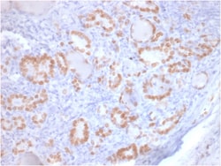
Biotium TTF-1 / NKX2.1 (Thyroid & Lung Epithelial Marker) (rNX2.1/690), CF568 conjugate, 0.1mg/mL
This antibody recognizes a protein of 40 kDa, identified as Thyroid transcription factor-1 (TTF-1). TTF-1 is a member of the NKx2 family of homeodomain transcription factors. It is expressed in epithelial cells of the thyroid gland and the lung. Nuclei from liver, stomach, pancreas, small intestine, colon, kidney, breast, skin, testes, pituitary, prostate, and adrenal glands are unreactive. Anti-TTF-1 is useful in differentiating primary adenocarcinoma of the lung from metastatic carcinomas originating in the breast, mediastinal germ cell tumors, and malignant mesothelioma. It can also be used to differentiate small cell lung carcinoma from lymphoid infiltrates. Loss of TTF-1 expression in non-small cell lung carcinoma has been associated with aggressive behavior of such neoplasms. TTF-1 reactivity is also seen in thyroid malignancies.Primary antibodies are available purified, or with a selection of fluorescent CF Dyes and other labels. CF Dyes offer exceptional brightness
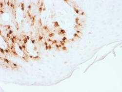
Biotium ARF1 (Golgi Apparatus Marker)(3F1), CF740 conjugate, 0.1mg/mL
The ADP-ribosylation factor (ARF) family comprises a group of structurally and functionally conserved proteins, which are members of the Ras superfamily of regulatory GTP-binding proteins. The ARF family is divided functionally into the ARF and the ARF-like proteins. ARF's share more than 60% sequence identity, appear to be ubiquitous in eukaryotes, and are highly conserved evolutionarily. ARF is involved in intracellular protein traffic to and within the Golgi complex. ARF has a number of disparate activities including maintenance of organelle integrity, assembly of coat proteins, as a co-factor for cholera toxin and as an activator of phospholipase D. Primary antibodies are available purified, or with a selection of fluorescent CF Dyes and other labels. CF Dyes offer exceptional brightness and photostability. Note: Conjugates of blue fluorescent dyes like CF405S and CF405M are not recommended for detecting low abundance targets, because blue dyes have lower fluorescenc

Biotium Rb1 (Tumor Suppressor Protein) (1F8), CF647 conjugate, 0.1mg/mL
This antibody recognizes a 105 kDa phosphoprotein, identified as retinoblastoma (Rb) gene product. Its epitope is localized between aa 703-772. It shows no cross reaction with p107 or p130. It specifically stains the nuclei of BT-20 cells and primary human foreskin fibroblast (HFF) cells. It does not stain the Rb-negative BT549 cells. It reacts with the hyperphosphorylated as well as the un (under) phosphorylated form of the Rb protein. Retinoblastoma gene product plays a key role in cell cycle control. It has been identified as a tumor suppressor gene whose loss of its function leads to tumor development. It is widely expressed in a variety of human tissues including breast, esophageal, squamous cell and cervical carcinoma.Primary antibodies are available purified, or with a selection of fluorescent CF Dyes and other labels. CF Dyes offer exceptional brightness and photostability. Note: Conjugates of blue fluorescent dyes like CF405S and CF405M are not recommended for d
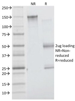
Biotium VEGF (Vascular Endothelial Growth Factor)(VG76e), 0.2mg/mL
This MAb recognizes proteins of 19-22 kDa (reducing) and 38 kDa-44 kDa (non-reducing), identified as various isoforms of Vascular Endothelial Growth Factor or Vascular Permeability Factor (VEGF/VPF). It is highly specific to VEGF, which is a homodimeric, disulfide-linked glycoprotein with a close homology to platelet-derived growth factor (PDGF). There are multiple isoforms of VEGF containing 206-, 189-, 165-, and 121-amino acid residues. The smaller two isoforms, VEGF165 and VEGF121, are secreted proteins and act as diffusible agents, whereas the larger two remain cell associated. VEGF/VPF plays an important role in angiogenesis, which promotes tumor progression and metastasis. Primary antibodies are available purified, or with a selection of fluorescent CF Dyes and other labels. CF Dyes offer exceptional brightness and photostability. Note: Conjugates of blue fluorescent dyes like CF405S and CF405M are not recommended for detecting low abundance targets, because blue d
Biotium Histone H1 (Nuclear Marker) (r1415-1), CF680 conjugate, 0.1mg/mL
Eukaryotic histones are basic and water-soluble nuclear proteins that form hetero-octameric nucleosome particles by wrapping 146 base pairs of DNA in a left-handed supealpha-helicalturn sequentially to form chromosomal fiber. Two molecules of each of the four core histones (H2A, H2B, H3, and H4) form the octamer; formed of two H2A-H2B dimers and two H3-H4 dimers, forming two nearly symmetrical halves by tertiary structure. Over 80% of nucleosomes contain the linker Histone H1, derived from an intronless gene that interacts with linker DNA between nucleosomes and mediates compaction into higher order chromatin. Histones are subject to posttranslational modification by enzymes primarily on their N-terminal tails, but also in their globular domains. Such modifications include methylation, citrullination, acetylation, phosphorylation, sumoylation, ubiquitination and ADP-ribosylation.Primary antibodies are available purified, or with a selection of fluorescent CF Dyes and other l
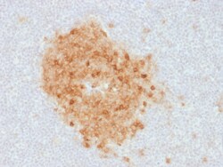
Biotium MART-1 / Melan-A / MLANA (Melanoma Marker) (MLANA/1761R), Biotin conjugate, 0.1mg/mL
This antibody recognizes a protein doublet of 20-22 kDa, identified as MART-1 (Melanoma Antigen Recognized by T cells 1) or Melan-A. MART-1 is a newly identified melanocyte differentiation antigen recognized by autologous cytotoxic T lymphocytes. Seven other melanoma associated antigens recognized by autologous cytotoxic T cells include MAGE-1, MAGE-3, tyrosinase, gp100, gp75, BAGE-1, and GAGE-1. Subcellular fractionation shows that MART-1 is present in melanosomes and endoplasmic reticulum. This MAb labels melanomas and other tumors showing melanocytic differentiation. It is also a useful positive-marker for angiomyolipomas. It does not stain tumor cells of epithelial, lymphoid, glial, or mesenchymal origin.Primary antibodies are available purified, or with a selection of fluorescent CF Dyes and other labels. CF Dyes offer exceptional brightness and photostability. Note: Conjugates of blue fluorescent dyes like CF405S and CF405M are not recommended for detecting low a
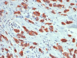
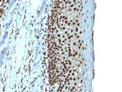
Biotium Prostate Specific Antigen (PSA)(3E6), Biotin conjugate, 0.1mg/mL
Recognizes a single protein of 33-34 kDa, identified as the prostate specific antigen (PSA). This MAb is highly specific to PSA and stains prostatic secretory and ductal epithelium in both normal and neoplastic tissues. PSA is a chymotrypsin-like serine protease (kallikrein family) exclusively produced by the prostate epithelium, and abundant in seminal fluid. PSA can be detected in the sera of patients with prostatic carcinoma. It is predominantly complexed to a liver-derived serine protease inhibitor, alpha-1-antichymotrypsin (ACT). A higher proportion of serum PSA is complexed to ACT in prostate cancer than in benign prostate hyperplasia. This MAb makes an excellent pair with MAb A67-B/E3 for PSA tests.Primary antibodies are available purified, or with a selection of fluorescent CF Dyes and other labels. CF Dyes offer exceptional brightness and photostability. Note: Conjugates of blue fluorescent dyes like CF405S and CF405M are not recommended for detecting low ab
Biotium Insulin Receptor Alpha(INSR/1661), CF740 conjugate, 0.1mg/mL
The insulin receptor (INSR) is a heterodimeric protein complex that has an intracellular subunit, which is disulfide-linked to a transmembrane segment. The insulin ligand binds to the INSR and initiates molecular signaling pathways that promote glucose uptake in cells and glycogen synthesis. Insulin binding to INSR induces phosphorylation of intra-cellular tyrosine kinase domains and recruitment of multiple SH2 and SH3 domain-containing intracellular proteins that serve as signaling intermediates for pleiotropic effects of insulin. Type 1 diabetes is an autoimmune condition of the endocrine pancreas that results in destruction of insulin secreting cells and a progressive loss in insulin-sensitive glucose uptake by cells. Primary antibodies are available purified, or with a selection of fluorescent CF Dyes and other labels. CF Dyes offer exceptional brightness and photostability. Note: Conjugates of blue fluorescent dyes like CF405S and CF405M are not recommended for dete
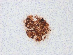
Biotium Insulin / IRDN (beta-Cell & Insulinoma Marker)(rIRDN/805), 1mg/mL
This antibody recognizes a polypeptide which is identified as insulin, a 51-amino acid polypeptide composed of A and B chains connected through the C-peptide. Proinsulin, which has very little biological activity, is cleaved by proteases within its cell of origin into the insulin molecule and the C-terminal basic residue. Insulin enhances membrane transport of glucose, amino acids, and certain ions. It also promotes glycogen storage, formation of triglycerides, and synthesis of proteins and nucleic acids. Deficiency of insulin results in diabetes mellitus. The main storage site for insulin is the pancreatic islets. Antibodies to insulin are important as beta-cell and insulinoma markers.Primary antibodies are available purified, or with a selection of fluorescent CF Dyes and other labels. CF Dyes offer exceptional brightness and photostability. Note: Conjugates of blue fluorescent dyes like CF405S and CF405M are not recommended for detecting low abundance targets, because